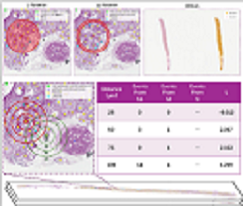
High-throughput ”pathomic” analysis of Whole Slide Images (WSIs) offers new opportunities to study tissue characteristics and for biomarker discovery. However, the clinical relevance of the tissue characteristics at the micro- and macro-environment level is limited by the lack of tools that facilitate the measurement of the spatial interaction of individual structure characteristics and their association with clinical parameters. To address these challenges, we introduce HistoWAS (Histology-Wide Association Study), a computational framework designed to link tissue spatial organization to clinical outcomes. Specifically, HistoWAS implements (1) a feature space that augments conventional metrics with 30 topological and spatial features, adapted from Geographic Information Systems (GIS) point pattern analysis, to quantify tissue micro-architecture; and (2) an association study engine, inspired by Phenome-Wide Association Studies (PheWAS), that performs mass univariate regression for each feature with statistical correction. As a proof of concept, we applied HistoWAS to analyze a total of 102 features (72 conventional object-level features and our 30 spatial features) using 385 PAS-stained WSIs from 206 participants in the Kidney Precision Medicine Project (KPMP). The code and data have been released to https: // github. com/ hrlblab/ histoWAS .
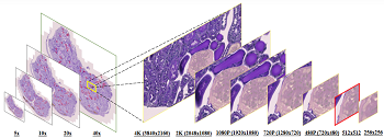
In digital pathology, the traditional method for deep learning-based image segmentation typically involves a two-stage process: initially segmenting high-resolution whole slide images (WSI) into smaller patches (e.g., 256 × 256, 512 × 512, 1024×1024) and subsequently reconstructing them to their original scale. This method often struggles to capture the complex details and vast scope of WSIs. In this paper, we propose the holistic histopathology (HoloHisto) segmentation method to achieve end-to-end segmentation on gigapixel WSIs, whose maximum resolution is above 80,000×70,000 pixels. HoloHisto fundamentally shifts the paradigm of WSI segmentation to an end-to-end learning fashion with 1) a large (4K) resolution base patch for elevated visual information inclusion and efficient processing, and 2) a novel sequential tokenization mechanism to properly model the contextual relationships and efficiently model the rich information from the 4K input. To our best knowledge, HoloHisto presents the first holistic approach for gigapixel resolution WSI segmentation, supporting direct I/O of complete WSI and their corresponding gigapixel masks. Under the HoloHisto platform, we unveil a random 4K sampler that transcends ultra-high resolution, delivering 31 and 10 times more pixels than standard 2D and 3D patches, respectively, for advancing computational capabilities. To facilitate efficient 4K resolution dense prediction, we leverage sequential tokenization, utilizing a pre-trained image tokenizer to group image features into a discrete token grid. To assess the performance, our team curated a new kidney pathology image segmentation (KPIs) dataset with WSI-level glomeruli segmentation from whole mouse kidneys. From the results, HoloHisto-4K delivers remarkable performance gains over previous state-of-the-art models.
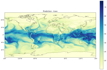
Compute-optimal scaling laws are relatively well studied for NLP and CV, where objectives are typically single-step and targets are comparatively homogeneous. Weather forecasting is harder to characterize in the same framework: autoregressive rollouts compound errors over long horizons, outputs couple many physical channels with disparate scales and predictability, and globally pooled test metrics can disagree sharply with per-channel, late-lead behavior implied by short-horizon training. We extend neural scaling analysis for autoregressive weather forecasting from single-step training loss to long rollouts and per-channel metrics. We quantify (1) how prediction error is distributed across channels and how its growth rate evolves with forecast horizon, (2) if power law scaling holds for test error, relative to rollout length when error is pooled globally, and (3) how that fit varies jointly with horizon and channel for parameter, data, and compute-based scaling axes. We find strong cross-channel and cross-horizon heterogeneity: pooled scaling can look favorable while many channels degrade at late leads. We discuss implications for weighted objectives, horizon-aware curricula, and resource allocation across outputs.
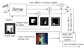
Full optical simulations of 3D scenes using commercial ray-tracing tools such as Speos provide physically accurate results but suffer from excessive computation time. To overcome this limitation, we propose a hybrid simulation pipeline that separates scene rendering and lens modeling into two stages. First, Zemax is used to precompute lens characteristics including distortion maps, depth-variant relative illumination, and a library of depth-dependent point spread functions (PSFs). Second, Speos is employed to render the ideal camera scene and per-pixel depth maps without optical degradation. Finally, a distributed multi-GPU system efficiently applies the Zemax-derived optical degradations to the Speos-rendered images. This approach significantly reduces simulation time while retaining the essential physical properties of lens degradation. The proposed framework provides realistic image formation data for testing computer vision and imaging system design.