
The most prominent problem in virtual reality (VR) technology is that users may experience motion-sickness-like symptoms when they immerse into a VR environment. These symptoms are recognized as visually induced motion sickness (VIMS) or virtual reality motion sickness. The objectives of this study were to investigate the association between the electroencephalogram (EEG) and subjectively rated VIMS level (VIMSL) and find EEG markers for VIMS evaluation. A VR-based vehicle-driving simulator was used to induce VIMS symptoms, and a wearable EEG device with four electrodes (the Muse) was used to collect EEG data. The results suggest that individual tolerance, susceptibility, and recoverability to VIMS varied largely among subjects; the following markers were shown to be significantly different from no-VIMS and VIMS states (P < 0.05): (1) means of gravity frequency (GF) for theta@FP1, alpha@TP9, alpha@FP2, alpha@TP10, and beta@FP1; (2) standard deviation of GF for alpha@TP9, alpha@FP1, alpha@FP2, alpha@TP10, and alpha@(FP2–FP1); (3) standard deviation of power spectral entropy for FP1; (4) means of Kolmogorov complexity (KC) for TP9, FP1, and FP2. These results also demonstrate that it is feasible to perform VIMS evaluation using an EEG device with a few electrodes.

Diabetic Retinopathy (DR) is a medical condition, also known as diabetic eye disease, which is vision-threatening damage to the retina of the eye caused by diabetes. As the technology advances, researchers are becoming more interested in intelligent medical diagnosis systems to assist screening of DR in earlier stages. In this study, variety of state-of-the-art procedures are used to extract the anatomic segments and lesions from the color fundus images. In addition, an automated system is proposed for the detection of anatomic segments and lesions by grading approach to help clinical diagnosis of the DR analysis. Four publicly available databases of color fundus images and various appropriate measurement techniques are used to compare quantitatively the performance of the proposed system. The experiments conducted on DIARETDB0, DIARETDB1, STARE, and HRF data sets have proved that accuracy, sensitivity, and specificity of the proposed system are comparable or superior to state-of-the-art methods.

Grating-based x-ray phase-contrast imaging has the potential to enhance image quality and provide inner structure details non-destructively. In this work, using grating-based x-ray phase-contrast imaging system and employing integrating-bucket method, the quantitative expressions of signal-to-noise ratios due to photon statistics and mechanical error are analyzed in detail. Photon statistical noise and mechanical error are the main sources affecting the image noise in x-ray grating interferometry. Integrating-bucket method is a new phase extraction method translated to x-ray grating interferometry; hence, its image quality analysis would be of great importance to get high-quality phase image. The authors’ conclusions provide an alternate method to get high-quality refraction signal using grating interferometer, and hence increases applicability of grating interferometry in preclinical and clinical usage.

Traditional spaghetti plots from ensemble data provide no explicit information as to the uncertainty of the realization flow paths. While intuitive assessment can be used when visualizing streamline density directly in such a plot, the display is often cluttered and difficult to interpret. The authors present a method to measure uncertainty and visualize member streamlines from an ensemble of vector fields. The method incorporates velocity probability density as a feature along each member streamline. The authors show visualizations of two different data sets using the proposed method.

A stereo matching algorithm is used to find the best match between a pair of images. To compute the cost of the matching points from the sequence of images, the disparity maps from video streams are estimated. However, the estimated disparity sequences may cause undesirable flickering errors. These errors result in low visibility of the synthesized video and reduce video coding. In order to solve this problem, in this article, the authors propose a spatiotemporal disparity refinement on local stereo matching based on the segmentation strategy. Based on segmentation information, matching point searching, and color similarity, adaptive disparity values to recover the disparity errors in disparity sequences can be obtained. The flickering errors are also effectively removed, and the boundaries of objects are well preserved. The procedures of the proposed approach consist of a segmentation process, matching point searching, and refinement in the temporal and spatial domains. Experimental results verify that the proposed approach can yield a high quantitative evaluation and a high-quality disparity map compared with other methods.

Three-dimensional (3D) reconstruction is extensively used in microscopic applications. Reducing excessive error points and achieving accurate matching of weak texture regions have been the classical challenges for 3D microscopic vision. A Multi-ST algorithm was proposed to improve matching accuracy. The process is performed in two main stages: scaled microscopic images and regularized cost aggregation. First, microscopic image pairs with different scales were extracted according to the Gaussian pyramid criterion. Second, a novel cost aggregation approach based on the regularized multi-scale model was implemented into all scales to obtain the final cost. To evaluate the performances of the proposed Multi-ST algorithm and compare different algorithms, seven groups of images from the Middlebury dataset and four groups of experimental images obtained by a binocular microscopic system were analyzed. Disparity maps and reconstruction maps generated by the proposed approach contained more information and fewer outliers or artifacts. Furthermore, 3D reconstruction of the plug gauges using the Multi-ST algorithm showed that the error was less than 0.025 mm.

Traditional approaches for the identification of leaf diseases involve the use of handcrafted features such as colors and textures for feature extraction. Therefore, these approaches may have limitations in extracting abundant and discriminative features. Although deep learning approaches have been recently introduced to overcome the shortcomings of traditional approaches, existing deep learning models such as VGG and ResNet have been used in these approaches. This indicates that the approach can be further improved to increase the discriminative power because the spatial attention mechanism to predict the background and spot areas (i.e., local areas with leaf diseases) has not been considered. Therefore, a new deep learning architecture, which is hereafter referred to as region-of-interest-aware deep convolutional neural network (ROI-aware DCNN), is proposed to make deep features more discriminative and increase classification performance. The primary idea is that leaf disease symptoms appear in leaf area, whereas the background region does not contain useful information regarding leaf diseases. To realize this, two subnetworks are designed. One subnetwork is the ROI subnetwork to provide more discriminative features from the background, leaf areas, and spot areas in the feature map. The other subnetwork is the classification subnetwork to increase the classification accuracy. To train the ROI-aware DCNN, the ROI subnetwork is first learned with a new image set containing the ground truth images where the background, leaf area, and spot area are divided. Subsequently, the entire network is trained in an end-to-end manner to connect the ROI subnetwork with the classification subnetwork through a concatenation layer. The experimental results confirm that the proposed ROI-aware DCNN can increase the discriminative power by predicting the areas in the feature map that are more important for leaf diseases identification. The results prove that the proposed method surpasses conventional state-of-the-art methods such as VGG, ResNet, SqueezeNet, bilinear model, and multiscale-based deep feature extraction and pooling.

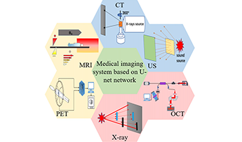
Medical image analysis is performed by analyzing images obtained by medical imaging systems to solve clinical problems. The purpose is to extract effective information and improve the level of clinical diagnosis. In recent years, automatic segmentation based on deep learning (DL) methods has been widely used, where a neural network can automatically learn image features, which is in sharp contrast with the traditional manual learning method. U-net is one of the most important semantic segmentation frameworks for a convolutional neural network (CNN). It is widely used in the medical image analysis domain for lesion segmentation, anatomical segmentation, and classification. The advantage of this network framework is that it can not only accurately segment the desired feature target and effectively process and objectively evaluate medical images but also help to improve accuracy in the diagnosis by medical images. Therefore, this article presents a literature review of medical image segmentation based on U-net, focusing on the successful segmentation experience of U-net for different lesion regions in six medical imaging systems. Along with the latest advances in DL, this article introduces the method of combining the original U-net architecture with deep learning and a method for improving the U-net network.

When portraits taken under various illuminants are placed together, they appear disparate from each other due to observers’ chromatic adaption to each portrait locally. Because the human perception has strong attachment to the memory color of human skin, it may cause an affective effect. This study intends to identify the affective effect of viewing multiple number of portraits whose white balance are not aligned. A visual assessment was conveyed, where portraits from different sources were matched. To simulate various illuminant conditions, three alterations of white balance were prepared for each portrait-warm (3000 K), neutral (6500 K), and cool tones (8000 K). This makes nine combinations for each stimulus. In Study I, 31 Korean college students were presented with a random one by one combination. Based on their subjective judgments on five criteria—authenticity, naturalness, professionality, content conveyance, and overall satisfaction—a statistical analysis was performed. In Study II, a creative writing session was followed in that the participants presumed the message of the portrait matches, and the content analysis was conducted. The results showed that a match of neutrally balanced portraits best appealed authentic and professional styles while minimizing any affective bias. When matches consisted of contrasting tones, on the other hand, they delivered content conveyance better while sacrificing naturalness of portraits. The white balance is limited to three types though this study provides evidence that it is a matter of choice to align, adjust, or alter the white balance of multi-sourced portraits depending on the aiming affective effect.