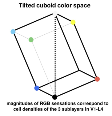
At HVEI-2012, I presented a neurobiologically-based model for trichromatic color sensations in humans, mapping the neural substrate for color sensations to V1-L4—the thalamic recipient layer of the primary visual cortex. In this paper, I propose that V1-L4 itself consists of three distinct sub-layers that directly correspond to the three primary color sensations: blue, red, and green. Furthermore, I apply this model to three aspects of color vision: the three-dimensional (3D) color solid, dichromatism, and ocular agnosticism. Regarding these aspects further: (1) 3D color solid: V1-L4 is known to exhibit a gradient of cell densities from its outermost layer (i.e., its pia side) to its innermost layer (i.e., its white matter side). Taken together with the proposition that the population size of a cell assembly directly corresponds with the magnitude of a color sensation, it can be inferred that the neurobiologically-based color solid is a tilted cuboid. (2) Chromatic color blindness: Using deuteranopia as an example, at the retinal level, M-cones are lost and replaced by L-cones. However, at the cortical level, deuteranopia manifests as a fusion of the two bottom layers of V1-L4. (3) Ocular agnosticism: Although color sensation is monocular, we normally are not aware of which eye we are seeing with. This visual phenomenon can be explained by the nature of ocular integration within V1-L4. A neurobiologically-based model for human color sensations could significantly contribute to future engineering efforts aimed at enhancing human color experiences.

Today, different models and instruments exist to study and model color vision and color vision deficiency. These systems are often modeled at spectral and retinal levels. In this study, we propose a novel approach to set up models and aids for color vision deficiencies, considering the role of spatial color processing in human visual system. In particular, we present the results of a perceptual test to identify the role of the spatial arrangement in color discrimination by Color Deficient Observers (CDOs) and Color Normal Observers (CNOs), using simultaneous contrast effect.

In the last 80 years, the role of spatial processing in the visual system has been analyzed and demonstrated from many studies and experiments. Starting from the first studies of Young, Helmholtz and Hering, color vision models have developed, and several biological and physiological research paper proved the importance of spatial processing in color vision. In this paper, we present some of the studies which have explored the role of spatial processing to study color vision deficiency. Main scope of this work is to increase the awareness of the scientific community on the importance to include spatial processing not only in color vision models, but also in developing color deficiency aids and tests.