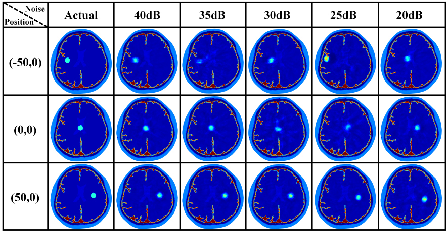
Cerebral hemorrhage is a common cerebrovascular disease, and magnetic induction tomography (MIT), a new electromagnetic imaging technique is used as a good auxiliary method for preoperative and postoperative bedside monitoring, and supplement computed tomography (CT) and magnetic resonance imaging (MRI) studies. In practical applications, space constraints limit the number of coils set up by the MIT system, which in turn limits the imaging information obtained from the detection coils. In order to reduce the number of coils while ensuring imaging accuracy and explore the relationship between the relative positions of coil arrays and lesions so as to obtain better reconstruction results, a regional detection method of MIT for cerebral hemorrhage based on the stacked autoencoder (SAE) neural network algorithm is proposed. Based on the complex brain model, simulation experiments were carried out to divide into different regions according to the position of the coils and compare the reconstruction effect of the lesions. Results showed that the reconstruction effect of the lesions near the excitation coil was poor, and the reconstruction effect of the lesions near the detection coil was better. Phantom experiments were carried out to further verify the results. The sensitive region detection approach provides a novel idea for MIT optimization in detecting cerebral hemorrhage.
Ruijuan Chen, Xinlei Zhu, Yalin Du, Zhuangzhuang Zhao, Hongsheng Sun, Huiquan Wang, "Optimization of Region Detection for Magnetic Induction Tomography of Cerebral Hemorrhage based on Stacked Autoencoder Algorithm" in Journal of Imaging Science and Technology, 2025, pp 1 - 10, https://doi.org/10.2352/J.ImagingSci.Technol.2025.69.4.040508
 Find this author on Google Scholar
 Find this author on PubMed