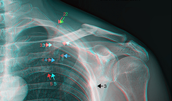
We present several stereoscopic 3D radiographs, obtained in a clinical setting using a technique requiring minimal operator training and no new technology. Reviewing known perceptual advantages in stereoscopic imaging, we argue the benefits for diagnosis and treatment planning primarily in orthopedics, with opportunities likely extended to rheumatology, oncology, and angiology (vascular medicine/surgery). These advantages accrue with the marginal additional cost of capturing just two or three supplementary radiographs using the proposed method. Presently, computed tomography (CT) scanning is standard for obtaining 3D imagery. We discuss relative advantages of stereoscopic 3D radiography (3DSR) in imaging resolution, cost, availability, and radiation dose. Further discussion will describe obstacles and challenges likely to be encountered in clinical implementation of 3DSR, to be mitigated through targeted training of clinicians and technicians. Further research is needed to explore and empirically validate the potential value of 3DSR. We hope to pave the way for this more accessible and cost-effective 3D imaging solution, enhancing diagnostic capabilities and treatment planning, especially in resource-constrained settings. (All 3D radiographs presented in this paper are in the red/cyan color anaglyph format. 3D anaglyph glasses are commonly available online, or in most comics bookstores. This report’s images can be found in L-R stereo-pair format, suitable for 3D viewing with a 3D screen or stereoscope, at this URL: https://www.starosta.com/3DSR/)