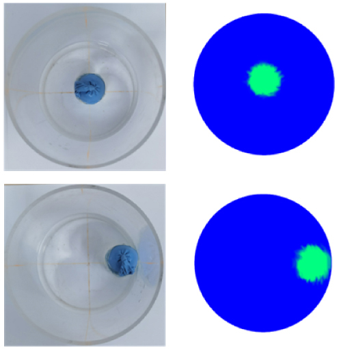
Magnetic induction tomography (MIT) is an emerging imaging technology holding significant promise in the field of cerebral hemorrhage monitoring. The commonly employed imaging method in MIT is time-difference imaging. However, this approach relies on magnetic field signals preceding cerebral hemorrhage, which are often challenging to obtain. Multiple bioelectrical impedance information with different frequencies is added to this study on the basis of single-frequency information, and the collected signals with different frequencies are identified to obtain the magnetic field signal generated by single-layer heterogeneous tissue. The Stacked Autoencoder (SAE) neural network algorithm is used to reconstruct the images of head multi-layer tissues. Both numerical simulation and phantom experiments are carried out. The results indicate that the relative error of the multi-frequency SAE reconstruction is only 7.82%, outperforming traditional algorithms. Moreover, under a noise level of 40 dB, the anti-interference capability of the MIT algorithm based on frequency identification and SAE is superior to traditional algorithms. This research explores a novel approach for the dynamic monitoring of cerebral hemorrhage and demonstrates the potential advantages of MIT in non-invasive monitoring.