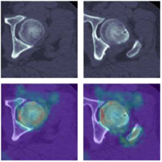
Computed tomography (CT) images provide a wealth of anatomical information crucial for diagnosing femoral fractures. However, predicting these fractures poses challenges due to postural variabilities of the femur and device-related factors. This study introduces an approach for predicting femoral fracture from CT and mask images. The approach includes several stages: annotations for masks, the scaling iterative closest point (SICP) algorithm for registration, three-dimensional (3D) affine transformation of images, image histogram matching, and a two-channel 3D convolutional neural network (3DCNN). In the proximal femoral region, SICP is applied to adjust the size and posture of the point cloud by using 3D affine transformation to ensure alignment with the target point cloud. The 3D affine transformation, generated by SICP registration, is applied to the original CT and mask images, systematically normalizing variances in the femoral postures and sizes across different subjects. Image histogram matching is used to diminish the variances in image grayscale values that originate from the scanning devices. It redistributes the pixel grayscale distributions in CT images, aligning them more closely with a reference histogram. The two-channel 3DCNN takes as input CT images (i.e., the first channel) that have undergone 3D affine transformation and image histogram matching, along with their corresponding masks (i.e., the second channel), and delivers the probability of a fracture as its output. Results show that the predictive capability of the 3DCNN-based model is notable, achieving an accuracy of 91.299%, specificity of 91.551%, sensitivity of 91.071%, and an area under the curve of 0.973. In conclusion, this approach effectively minimizes the impact of irrelevant factors on prediction, optimally utilizing image information to assess the risks of femoral fracture. Moreover, this approach enhances the accuracy and reliability of fracture prediction.